Avatar photo

About Dr. rer. nat. Dagmar Dietrich

Chemnitz University of Technology, Institute of Materials Science and Engineering, Chair of Surface Technology/Functional Materials, 09107 Chemnitz, Germany

A new insight into the phosphorus distribution of nanocrystalline Ni-Ni3P-diamond composites

The microstructure of an electroplated Ni-Ni3P-diamond composite has been studied by field-emission scanning electron microscopy, energy dispersive X-ray spectrometry and transmission Kikuchi diffraction. The use of an electron transparent sample reduced the resolution limits of X-ray spectrometry and electron backscatter diffraction. Basing on the P distribution and Ni/Ni3P orientation maps, standard observations made by backscattered electron imaging can be easily interpreted.


Microstructure and Particle Incorporation Behavior of Electrocodeposited Ni-Al2O3 Nanocomposites

Nickel-alumina composite films were obtained by electrocodeposition using different deposition techniques, viz. direct current (DC) deposition and pulse-reverse plating (PRP). Particle incorporation was determined by means of energy-dispersive X-ray spectroscopy and glow discharge optical emission spectrometry (GD-OES). The structure of the films was analyzed using electron microscopy, viz. scanning electron microscopy (SEM), electron backscatter diffraction (EBSD), transmission electron microscopy (TEM), and X-ray diffraction. A <100> fiber texture was found for pure nickel films, which was reduced due to a change in plating conditions and particle incorporation. EBSD mappings indicate that the nanosized particles inhibit nickel growth and thus lead to a smaller nickel crystallite size combined with a distinct loss of the <100> texture. Scanning transmission electron microscopy (STEM) and TEM reveal that the inclusion of alumina nanoparticles preferentially takes place in the grain boundary region where the particles terminate the growth of nickel. High-resolution TEM imaging proves a complete embedding of nanoparticles by the nickel matrix without any voids.


Anodic Film Formation in Oxalic Acid on AlMn0.5Mg0.5 Alloy

The microstructure of oxalic-acid-anodized layers on AlMn0.5Mg0.5 alloy is compared to such layers on aluminum. Differences originate from four types of precipitates occurring in the alloy, forming inclusions in the layers, roughening surface and interface, and modifying typical pore structures of anodized aluminum oxides. A characteristic feature of this modification is the appearance of transverse channels in pore walls. Nanoscaled precipitates are suggested as their origin. Correlation to functional properties such as microhardness and electrical isolation behavior is discussed.


Properties of Gold Composites with Nanostructured Carbon-based Materials

Results of electrocodeposition of gold matrix composite coatings with carbon-based materials are reported, namely ultradispersed diamonds (UDD) and multiwalled carbon nanotubes (MWCNT). Pure gold and gold composite coatings were prepared from a gold sulphite electrolyte with bath loads from 5 to 20 g/l (UDD bath) and from 0.1 to 5 g/l (MWCNT bath). The resulting composites are characterized in terms of carbon content, particle distribution, and their bonding to the matrix, surface morphology, and the influence of particle loading in the electrolyte on matrix microstructure. Vickers hardness, friction, and wear behavior were investigated and are discussed in terms of microstructure characterization. Some notable improvements in the performance of the composites were observed with regard to application as sliding contacts.


Strontium-Substituted Hydroxyapatite Coatings on Titanium by Electrodeposition Technique

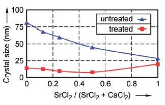
For the first time, strontium hydrogen phosphate (SrHPO4) was electrocrystallized on titanium substrate by means of electrochemical deposition technique, and converted to strontium hydroxyapatite (Sr10(PO4)6(OH)2) to improve implant adhesion and bone mineralization. Brushite (calcium phosphate dihydrate CaHPO4·2H2O) and strontium hydrogen phosphate were co-electrocrystallized on titanium substrate. With increasing SrCl2 and decreasing CaCl2 in the solution, Sr concentration in the coating was increased. Calcium substitution by strontium ranged from 0 to 100 atomic percent, thus having significant effect on layer thickness, morphology, and composition. Layers containing brushite and strontium hydrogen phosphate were converted to calcium hydroxyapatite and strontium hydroxyapatite. Strontium hydroxyapatite was formed in the case of 100 percent SrCl2 substituting CaCl2. Surface morphology, chemical composition, and phase identification of the coatings were studied by scanning electron microscopy combined with energy dispersive spectrometry (SEM-EDXS) and by X-ray diffractometry (XRD). Effects of the varying Sr substitution on the microstructure and properties are discussed.