Avatar photo

About Prof. Dr.-Ing. habil. Thomas Lampke

Chair of Surface Technology/Functional Materials, Chemnitz University of Technology, 09107 Chemnitz, Germany, Email: thomas.lampke@mb.tu-chemnitz.de

Please find bellow all articles from this author which were published in our journal. Lists with articles the author published in our or other journals, can be found at the following databases:

ResearcherID
Not listed
TEMA Database
German articles
English articles
ORCID
Not listed

Pulse plating of nickel-based alloys

Fig. 1: Cathodic polarisation curves for (a) Ni-Co, (b) Sn-Ni and (c) Ni-W electrolyte systems recorded at a scan rate of 10mVs-1 on a rotating disk electrode at different electrode rotating velocity: 0, 100, 600 and 1000rpm

In the present work, nickel-based alloy coatings (Ni-Co, Sn-Ni and Ni-W) with different microstructures were produced under direct and pulse current conditions. These alloys are of interest as potential replacement for chromium based coatings, especially hard chromium. A replacement for hard chromium coating faces the challenge of providing sufficient hardness values.  For some sliding wear applications, hardness might not be required at the same level as hard chromium, and coating toughness might be more critical.
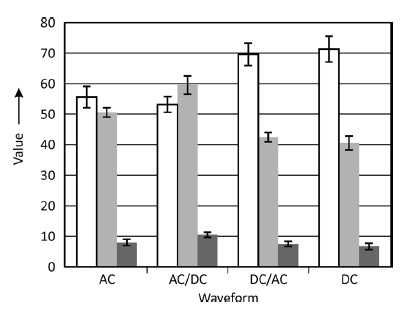
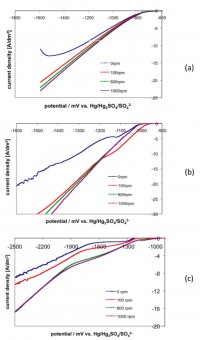
The effects of the pulse parameters (pulse waveform, pulse frequency and average current density) on the deposit structure and properties of these three systems have been investigated. Pulses have been defined based on the results of electrochemical measurements and numerical process simulation. The surface morphology, microstructure and microhardness of the deposit have been correlated to the pulse parameters applied.
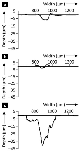
The experimental results showed that applying pulse plating substantially altered the properties of the coatings. The resulting layers exhibited a nano-crystalline microstructure, improved layer compactness and hardness of the nickel-based alloy deposits.


A new insight into the phosphorus distribution of nanocrystalline Ni-Ni3P-diamond composites

The microstructure of an electroplated Ni-Ni3P-diamond composite has been studied by field-emission scanning electron microscopy, energy dispersive X-ray spectrometry and transmission Kikuchi diffraction. The use of an electron transparent sample reduced the resolution limits of X-ray spectrometry and electron backscatter diffraction. Basing on the P distribution and Ni/Ni3P orientation maps, standard observations made by backscattered electron imaging can be easily interpreted.


Microstructure and Particle Incorporation Behavior of Electrocodeposited Ni-Al2O3 Nanocomposites

Nickel-alumina composite films were obtained by electrocodeposition using different deposition techniques, viz. direct current (DC) deposition and pulse-reverse plating (PRP). Particle incorporation was determined by means of energy-dispersive X-ray spectroscopy and glow discharge optical emission spectrometry (GD-OES). The structure of the films was analyzed using electron microscopy, viz. scanning electron microscopy (SEM), electron backscatter diffraction (EBSD), transmission electron microscopy (TEM), and X-ray diffraction. A <100> fiber texture was found for pure nickel films, which was reduced due to a change in plating conditions and particle incorporation. EBSD mappings indicate that the nanosized particles inhibit nickel growth and thus lead to a smaller nickel crystallite size combined with a distinct loss of the <100> texture. Scanning transmission electron microscopy (STEM) and TEM reveal that the inclusion of alumina nanoparticles preferentially takes place in the grain boundary region where the particles terminate the growth of nickel. High-resolution TEM imaging proves a complete embedding of nanoparticles by the nickel matrix without any voids.


Sol-Gel Coating for Aluminum Alloy: Self-Healing Characteristics

Water-based sol-gel coatings have been developed in the last years as cost-efficient and environmentally friendly protective layers. The stability of sol-gel films is often limited in the absence of functional compounds to generate self-healing. In this work, a water-based sol-gel coating was applied on aluminum alloy 6082. Benzotriazole and cerium nitrate were used as dopants and added to the sol after aging. The coating was injured in order to generate self-healing. The self-repairing feature of the defected specimen was evaluated by electrochemical impedance spectroscopy (EIS) in chloride solution. Healing qualification was examined by monitoring the defect size during the immersion test. The experiments show that the samples coated with an inhibitor-doped film are better protected against corrosion even after long-time immersion in chloride solution. The defect size after healing was smaller than that of the samples coated with non-doped film, which exhibited low stability in chloride solution. The defect size was several times bigger after the corrosion test. A contact stylus instrument and a scanning electron microscope (SEM) were used to characterize the defect surface profiles before and after healing.


Anodic Film Formation in Oxalic Acid on AlMn0.5Mg0.5 Alloy

The microstructure of oxalic-acid-anodized layers on AlMn0.5Mg0.5 alloy is compared to such layers on aluminum. Differences originate from four types of precipitates occurring in the alloy, forming inclusions in the layers, roughening surface and interface, and modifying typical pore structures of anodized aluminum oxides. A characteristic feature of this modification is the appearance of transverse channels in pore walls. Nanoscaled precipitates are suggested as their origin. Correlation to functional properties such as microhardness and electrical isolation behavior is discussed.